Metastasis to lung
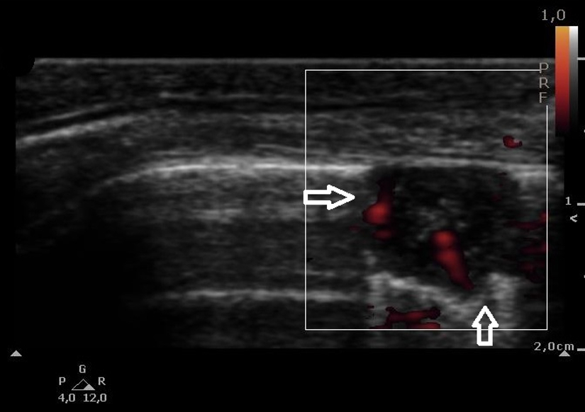
post by Natalia BudaColorectal cancer metastasis to lung. An oval subpleural consolidation marked by non-homogeneous echogenicity and echotexture, with disorganised vessels seen in PD (→↑). Linear transducer.
Neoplastic lesions are often marked by non-homogeneous echogenicity and echotexture. Flow patterns in both CD and PD are disorganised/chaotic. The shape can be regular (which is more often the case for metastatic lesions) or irregular (more frequent in the case of primary lesions). The pleural line is sometimes affected by the neoplastic process and this is when we can observe its fragmentariness, or even regional absence or a reduction of lung sliding caused by the infiltration of the chest wall. Often tumours located proximally in the bronchial tree (e.g. bronchogenic cancer) cause distal atelectasis: it is a resorption atelectasis, i.e. one caused by the cut-off of the airflow to distal parts of the bronchial tree.


