Pneumonia
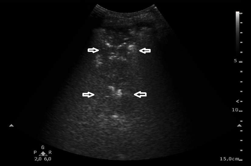
post by Natalia BudaPneumonia. A wedge-shaped consolidation with irregular margins and approx. 70 mm diameter (←→), with dynamic air bronchogram (numerous echoes against consolidations). Convex transducer.
Just like atelectasis, inflammatory lesions presented as a consolidation have a normally maintained flow in colour Doppler (CD) and power Doppler (PD) sonography. Additionally, a dynamic air bronchogram is seen within the consolidation – in other words, there is air in the bronchial tree (numerous echoes within the consolidation), which is visible during inspiration and vanishes partially or fully during expiration.